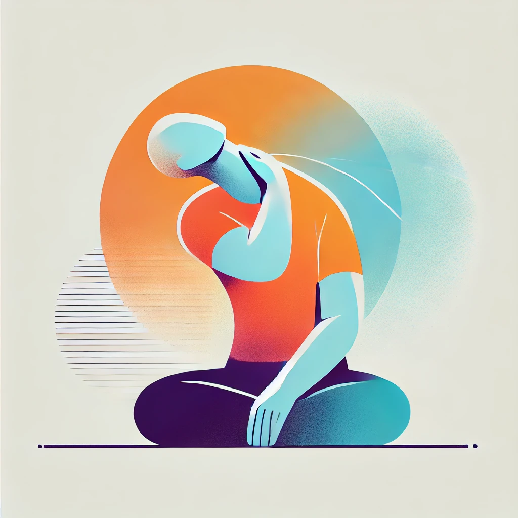
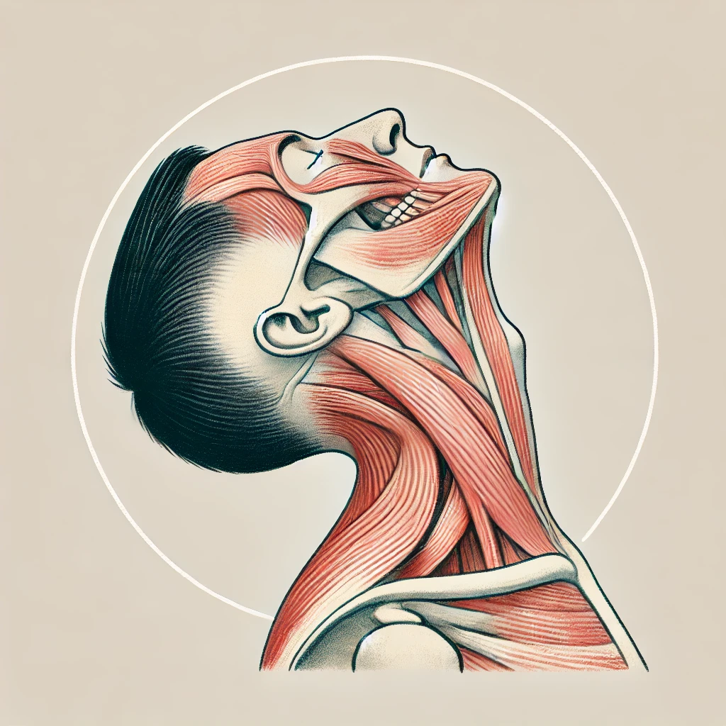
The oblique neck stretch is a neck exercise that helps improve flexibility and range of motion in the neck muscles. This exercise targets the scalene and other lateral neck muscles, which play a vital role in neck flexion, rotation, and lateral bending.
Regularly performing diagonal neck stretches can significantly improve flexibility and reduce neck discomfort. It is recommended to incorporate these exercises into your daily routine, especially if your job is sedentary or you engage in activities that put strain on your neck muscles.
Stand in the position shown in the image and get ready to perform the exercise.
The goal is to perform 2–4 repetitions on each side.
This stretch directly targets the neck muscles responsible for flexion, rotation, and lateral bending. By stretching these muscles regularly, you can increase your neck’s range of motion, making it easier to turn your head and look up or down without strain.
Tightness in the neck muscles can lead to pain and discomfort. Diagonal neck stretching helps release and lengthen these muscles, which can relieve existing pain and help prevent tension headaches or future neck pain. Research has shown that both static and diagonal stretches are effective in reducing neck-related disabilities and increasing neck range of motion.
Poor posture, especially forward head posture, can strain the neck muscles. Diagonal neck stretching promotes proper spinal alignment and stretches the muscles that help maintain correct posture. This can improve overall body mechanics and reduce the risk of pain in other areas, such as the lower back.
The neck and shoulder area are common sites for stress-related tension. Neck rotation stretches help relax these muscles, promoting a sense of calm and potentially reducing stress levels.
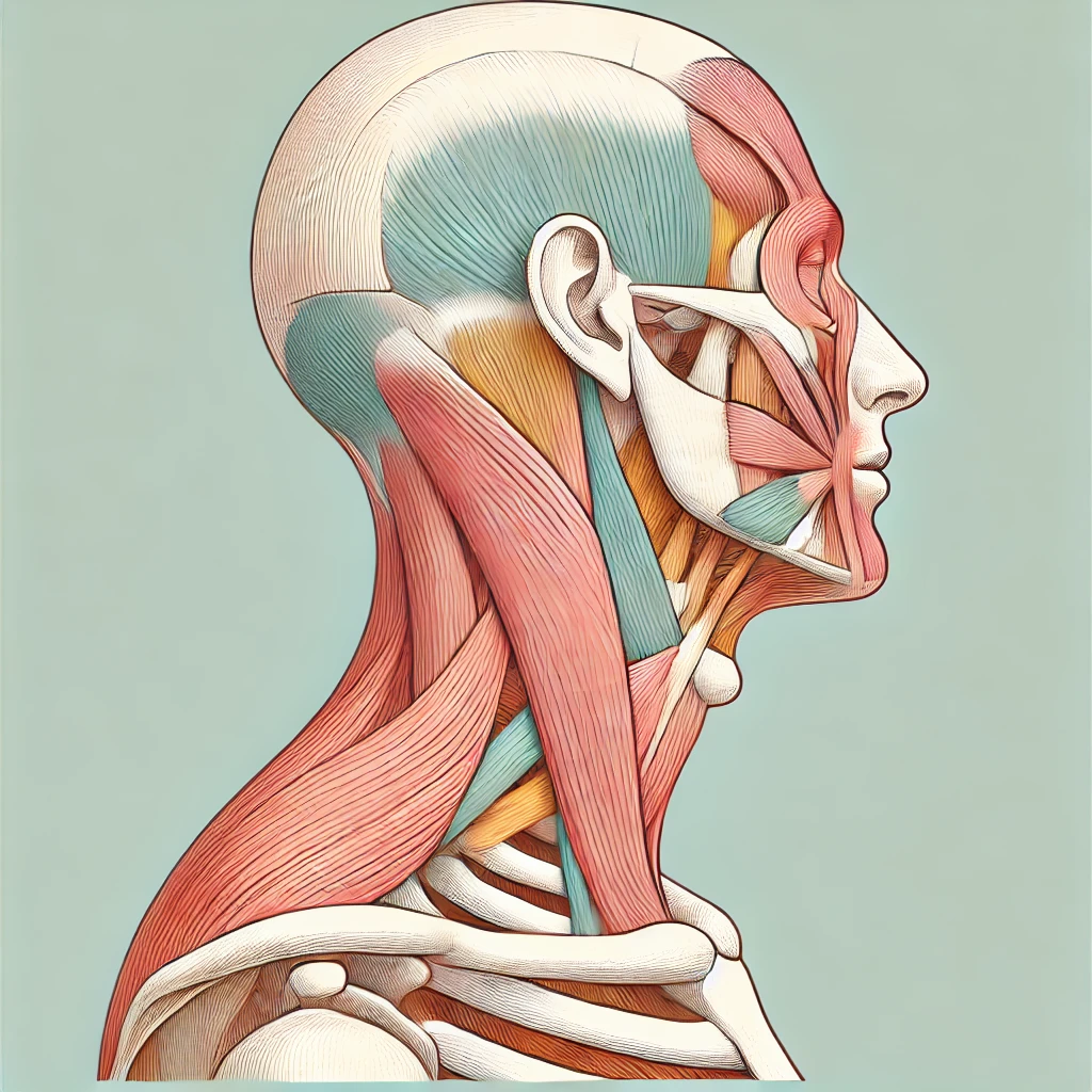
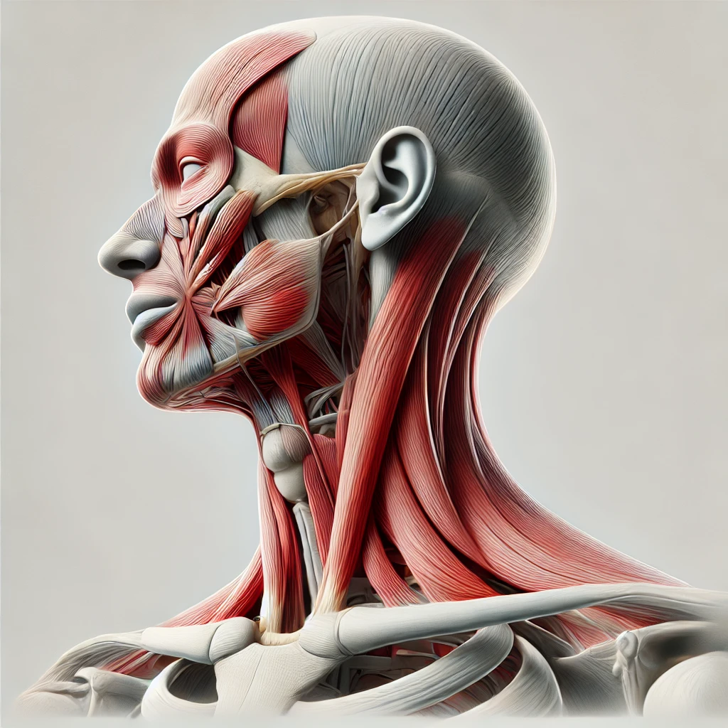
The levator scapulae is a long, slender muscle located in the neck and shoulder region, considered part of the deep neck muscles. It originates from the transverse processes of the cervical vertebrae (C1 to C4) and attaches to the medial border of the scapula near the superior angle.
The scalene muscles consist of three pairs (anterior, middle, and posterior) located on each side of the neck. These muscles assist in various movements, including lateral flexion of the neck, elevation of the first two ribs during inhalation, and rotation of the neck.
This muscle is a part of the longissimus, which belongs to the erector spinae muscle group. The longissimus capitis is located in the neck and head region and particularly assists in head rotation and extension. It originates from the upper neck and lower spine and attaches to the bones at the back of the head.
The longissimus cervicis is a muscle that is part of the erector spinae group and is located in the neck region. It originates from the upper thoracic vertebrae and attaches to the upper cervical vertebrae. Its main functions are assisting in neck extension (straightening the neck), lateral rotation, and maintaining the stability of the cervical spine. This muscle helps support and facilitate proper neck movement.
The semispinalis capitis is a deep, long muscle located in the neck and back of the head. It connects to the upper cervical and upper thoracic vertebrae and attaches to the bones of the skull, particularly the occipital and temporal regions.
The primary functions of the semispinalis capitis include extending the head backward and rotating it to the sides. In addition, this muscle helps stabilize the neck and maintain spinal alignment. It also plays a role in twisting movements of the neck and head during activities such as looking around or turning the head.
The semispinalis cervicis is a deep muscle in the neck region that originates from the upper part of the spine (T1–T6 vertebrae) and attaches to the upper cervical vertebrae (C2–C5). This muscle plays a key role in neck extension, rotation, and lateral flexion. It also helps stabilize and support the cervical spine and contributes to maintaining proper neck and spinal alignment.
The spinalis cervicis is a muscle that is part of the erector spinae group and is located in the neck region. It originates from the upper thoracic vertebrae (T2–T4) and attaches to the upper cervical vertebrae (C2). The spinalis cervicis is responsible for extending the neck and maintaining the upright position of the cervical spine. This muscle plays a role in strengthening and stabilizing the cervical spine and contributes to improved head and neck movements.
The spinalis capitis is a muscle that is part of the erector spinae group and is located in the head region. It originates from the upper cervical and thoracic vertebrae and attaches to the skull bones, particularly the occipital bone. The spinalis capitis is responsible for extending the head and assisting in maintaining the stability and support of the cervical spine and head.
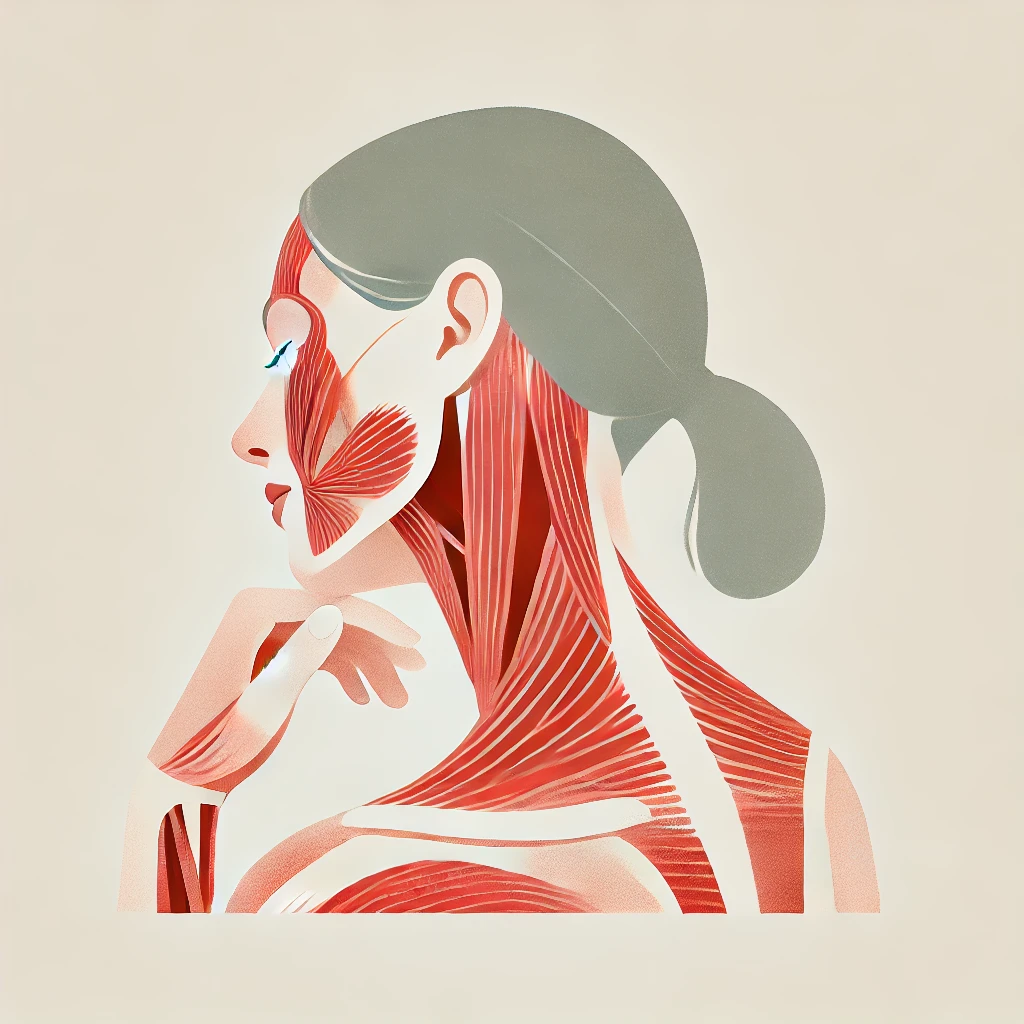
The sternocleidomastoideus is a large muscle in the neck that originates from the sternum and clavicle and attaches to the posterior part of the temporal bone (mastoid process). This muscle is responsible for rotating and flexing the neck and plays a role in lateral and forward neck movements. It also helps support and stabilize the head.
The trapezius is a large muscle in the back and neck region, shaped like a trapezoid, extending from the neck to the mid-back. It is composed of different parts and is divided into three main sections:
The trapezius muscle plays a key role in various shoulder and neck movements, including elevating the shoulders, rotating the scapulae, and moving the head backward. It also contributes to maintaining and stabilizing the shoulder and neck region.