


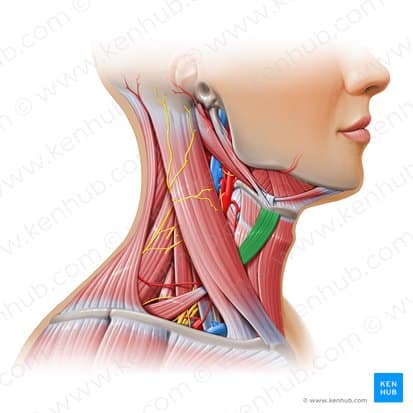


The neck muscles are among the most complex and vital parts of the body, playing a crucial role in supporting the head, facilitating its movements, and protecting essential pathways such as the spinal cord, blood vessels, and airways. These muscles are important not only for everyday motions but also for injury prevention and enhancing athletic performance. The intricate structure of the neck consists of multiple layers of muscles, nerves, and vessels that work together precisely. Understanding these structures helps us appreciate the importance of prevention, rehabilitation, and strengthening of these muscles. This article provides a comprehensive overview of the neck muscles, their functions, common injuries, and related training methods, offering valuable information for athletes, coaches, and the general public.
These muscles are primarily responsible for the major movements and stability of the neck.
The platysma is a thin, superficial muscle located at the front of the neck, extending from the upper chest to the lower jaw and facial skin. It plays a role in facial expressions, such as pulling the lower lip and wrinkling the skin of the neck, and indirectly helps prevent sagging of the neck skin.
Innervation: Provided by the cervical branch of the facial nerve (Facial Nerve).
Functions:
The platysma is an important focus in neck and facial cosmetic surgeries, and strengthening exercises for this muscle can help reduce sagging of the neck skin.
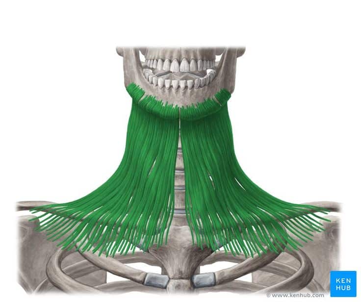
The sternocleidomastoid is a long, powerful muscle located on both sides of the neck. It originates from the sternum and clavicle and attaches to the mastoid process of the temporal bone. This muscle assists in the movements of the neck and head.
Innervation: Supplied by the accessory nerve (Cranial Nerve XI).
Functions:
This muscle plays a vital role in head and neck movements and is essential for maintaining balance and body coordination. Weakness or injury in this muscle can lead to restricted movement or neck pain.
These muscles lie beneath the superficial structures and play a role in stabilizing the neck and enabling more precise movements.
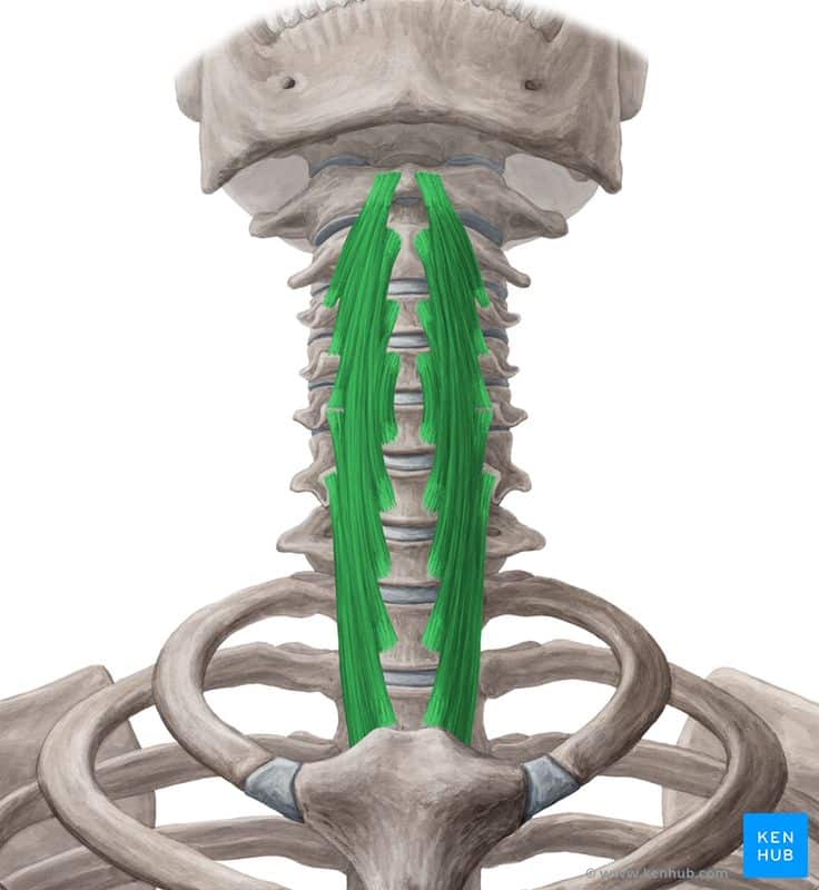
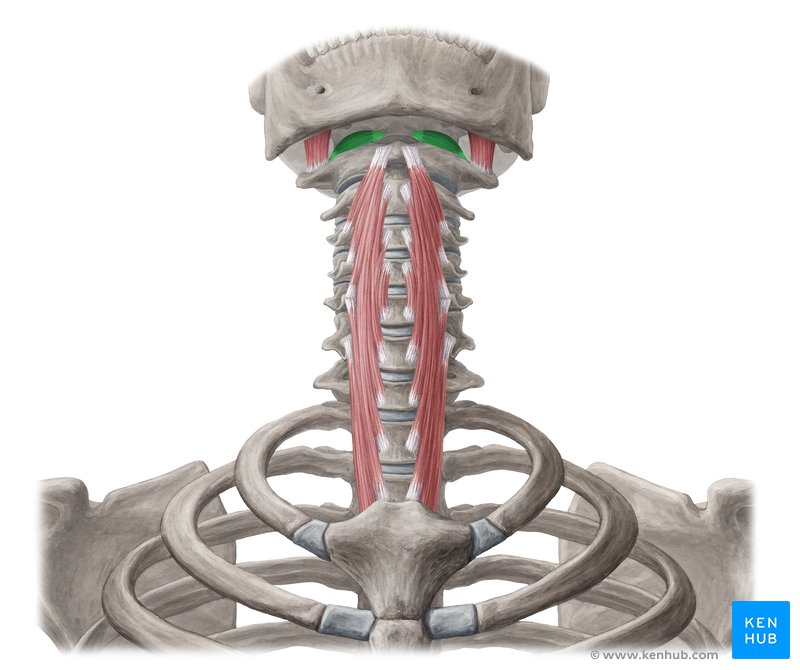
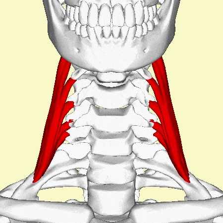
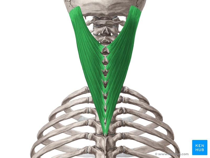
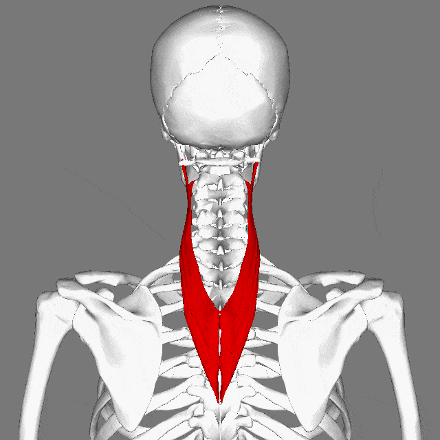
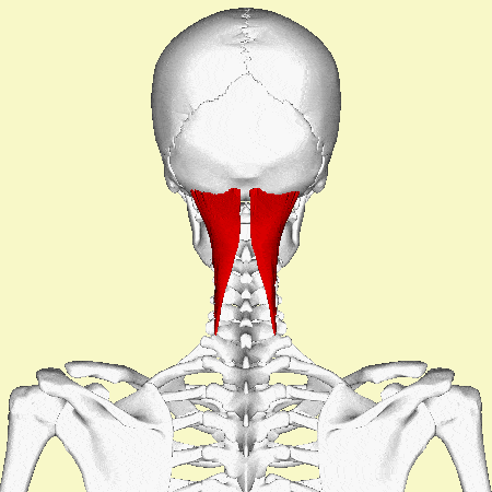
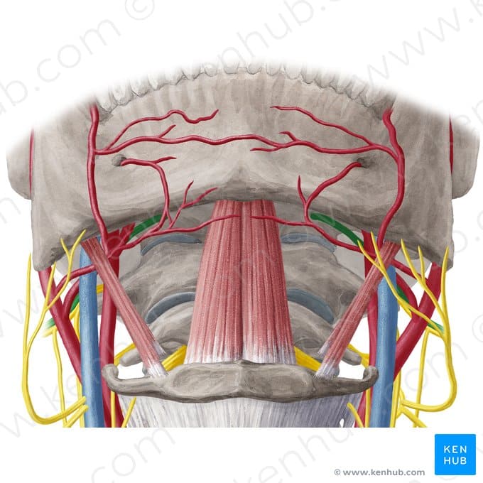
The longus colli is a deep muscle located in front of the cervical spine. It originates from the cervical vertebrae and some upper thoracic vertebrae, attaching to the transverse processes of other cervical vertebrae. This muscle is composed of three parts: superior, vertical, and inferior.
Innervation: Supplied by cervical spinal nerve branches (C2 to C6).
Functions:
This muscle plays a key role in stabilizing and controlling fine neck movements, and strengthening it can help improve posture and reduce neck pain.
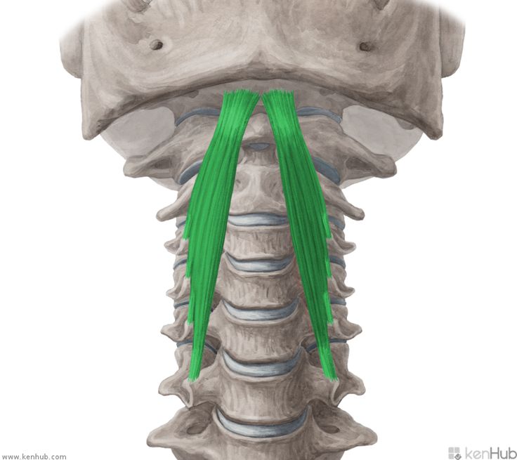
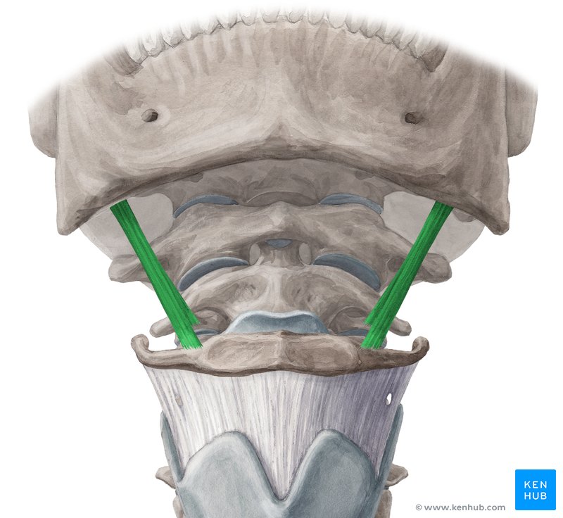
The longus capitis is a deep muscle located in front of the cervical spine. It originates from the transverse processes of cervical vertebrae C3 to C6 and attaches to the basilar part of the occipital bone. This muscle plays a role in head and neck movements.
Innervation: Supplied by cervical nerve branches (C1 to C3).
Functions:
This muscle plays an important role in maintaining posture and reducing neck tension by supporting fine neck movements and stabilizing the head. Strengthening it can help alleviate neck pain and improve neck stability.
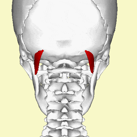
The rectus capitis anterior is a small, deep muscle located at the front of the cervical spine near the base of the skull. It originates from the transverse process of the atlas (C1) and attaches to the base of the occipital bone.
Innervation: Supplied by the anterior branches of the cervical spinal nerves (C1 and C2).
Functions:
This muscle plays a crucial role in small movements and stabilization of the skull, and strengthening or maintaining its health is important for improving neck posture and reducing pain caused by weakness in the deep neck muscles.
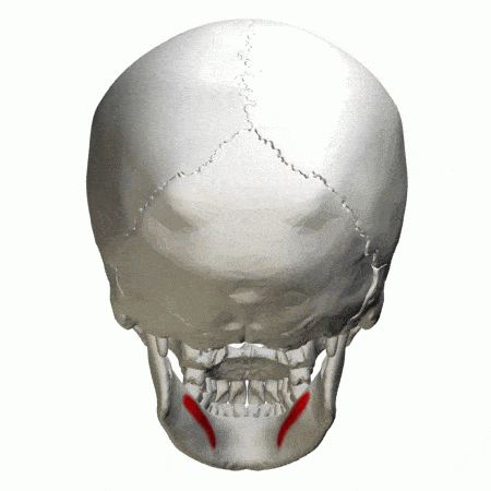
The rectus capitis lateralis is a small, deep muscle located on the sides of the cervical spine, playing a role in connecting the head to the neck. It originates from the transverse process of the atlas (C1) and attaches to the inferior surface of the occipital bone.
Innervation: Supplied by the anterior branches of the cervical spinal nerves (C1 and C2).
Functions:
This muscle plays an important role in controlled head movements and stability, helping to maintain proper alignment between the head and neck. Weakness or tension in this muscle can lead to impaired neck motion or cause neck pain.
These muscles play a role in lateral neck movements and assist with breathing.
The levator scapulae is a deep muscle of the neck and shoulder region that originates from the transverse processes of cervical vertebrae C1 to C4 and attaches to the superior part of the medial border of the scapula.
Innervation:
Supplied by branches of the cervical spinal nerves (C3 and C4) as well as the dorsal scapular nerve originating from the C5 nerve root.
Functions:
Clinical Importance:
This muscle can become tight or strained in conditions such as poor posture (e.g., prolonged sitting at a desk), which may lead to neck and shoulder pain. It is also involved in syndromes related to neck and upper scapular pain, such as myofascial pain syndrome.
The anterior scalene is a deep neck muscle that originates from the transverse processes of cervical vertebrae C3 to C6 and attaches to the first rib.
Innervation: Supplied by the anterior branches of the cervical spinal nerves (C4 to C6).
Functions:
This muscle plays an important role in neck movement and stability and can contribute to conditions such as thoracic outlet syndrome by compressing blood vessels and nerves.
The scalene medius is the largest muscle in the scalene group, originating from the transverse processes of cervical vertebrae C2 to C7 and attaching to the first rib.
Innervation: Supplied by the anterior branches of the cervical spinal nerves (C3 to C8).
Functions:
This muscle plays a role in lateral neck movements and breathing and may experience tension or compression in conditions such as thoracic outlet syndrome.
The posterior scalene is the smallest muscle in the scalene group. It originates from the transverse processes of cervical vertebrae C4 to C6 and attaches to the outer surface of the second rib.
Innervation: Supplied by the anterior branches of the cervical spinal nerves (C6 to C8).
Functions:
This muscle plays an important role in lateral neck movements and assists in the breathing process, and compared to other scalene muscles, it is less prone to compression or strain.
These muscles are responsible for rotational movements, extension (backward bending), and stabilization of the head and neck.
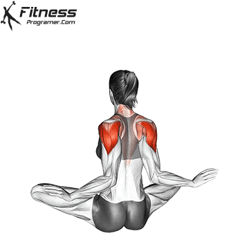
The trapezius is a large, superficial muscle located in the back, extending from the base of the skull to the thoracic vertebrae and attaching to the shoulders.
Innervation: Supplied by the accessory nerve (Cranial Nerve XI) and cervical nerve branches (C3 and C4).
Functions:
This muscle plays a vital role in shoulder and neck movements and contributes to the stability and balance of the upper body.
The splenius capitis is a superficial muscle located at the back of the neck. It originates from the cervical and upper thoracic vertebrae and attaches to the occipital bone and the mastoid process.
Innervation: Supplied by the dorsal rami of cervical spinal nerves (C3 and C4).
Functions:
This muscle plays a key role in rotational movements and extension of the head and neck, helping to maintain balance and alignment of the neck.
The splenius cervicis is a superficial muscle located at the back of the neck and upper back. It originates from the upper thoracic vertebrae (T3 to T6) and attaches to the transverse processes of cervical vertebrae C1 to C3.
Innervation: Supplied by the dorsal rami of cervical spinal nerves (C4 to C8).
Functions:
This muscle assists in neck movements and stability and plays a role in coordinating head and neck motions.
The semispinalis capitis is a deep muscle located at the back of the neck and upper back. It originates from the transverse processes of cervical and thoracic vertebrae (C4 to T6) and attaches to the posterior surface of the occipital bone.
Innervation: Supplied by the dorsal rami of the cervical spinal nerves.
Functions:
This muscle plays a key role in fine movements and stability of the head and neck.
The semispinalis cervicis is a deep muscle located at the back of the neck. It originates from the transverse processes of thoracic vertebrae (T1 to T6) and attaches to the spinous processes of cervical vertebrae (C2 to C5).
Innervation: Supplied by the dorsal rami of the spinal nerves.
Functions:
This muscle plays a key role in neck movement and stability and helps maintain proper neck posture.
The rectus capitis posterior major is a small, deep muscle located at the back of the neck. It originates from the spinous process of the axis (C2) and attaches to the inferior surface of the occipital bone.
Innervation: Supplied by the dorsal ramus of the first cervical spinal nerve (C1 – suboccipital nerve).
Functions:
This muscle plays an important role in fine movements and stabilization of the skull, helping to maintain head balance.
The rectus capitis posterior minor is a small, deep muscle located at the back of the neck. It originates from the posterior tubercle of the atlas (C1) and attaches to the inferior surface of the occipital bone.
Innervation: Supplied by the dorsal ramus of the first cervical spinal nerve (C1 – suboccipital nerve).
Functions:
This muscle plays an important role in small movements and stabilization of head posture.
The obliquus capitis superior is a small, deep muscle located at the back of the neck. It originates from the transverse process of the atlas (C1) and attaches to the inferior surface of the occipital bone.
Innervation: Supplied by the dorsal ramus of the first cervical spinal nerve (C1 – suboccipital nerve).
Functions:
This muscle plays a role in fine movements and stability of the head and is part of the suboccipital muscle group.
The obliquus capitis inferior is a small but strong muscle located at the back of the neck. It originates from the spinous process of the axis (C2) and attaches to the transverse process of the atlas (C1).
Innervation: Supplied by the dorsal ramus of the first cervical spinal nerve (C1 – suboccipital nerve).
Functions:
This muscle is the only suboccipital muscle that does not attach to the occipital bone, and its primary role is in head rotation.
These muscles play a role in swallowing and neck stabilization and are divided into two groups.
The mylohyoid is a thin, broad muscle forming the floor of the mouth. It originates from the inner surface of the mandible and attaches to the hyoid bone.
Innervation: Supplied by the mylohyoid nerve, a branch of the inferior alveolar nerve originating from the trigeminal nerve (cranial nerve V).
Functions:
This muscle plays an important role in swallowing, chewing, and tongue movements, forming the floor of the mouth.
The geniohyoid is a small, narrow muscle located in the floor of the mouth. It originates from the mental spines on the inner surface of the mandible and attaches to the hyoid bone.
Innervation: Supplied by a branch of the hypoglossal nerve (Cranial Nerve XII) originating from C1.
Functions:
This muscle plays an important role in swallowing and stabilizing the hyoid bone, contributing to oral and pharyngeal functions.
The stylohyoid is a thin, long muscle that originates from the styloid process of the temporal bone and attaches to the hyoid bone.
Innervation: Supplied by a branch of the facial nerve (Cranial Nerve VII).
Functions:
This muscle plays a key role in the swallowing process and stabilization of the hyoid bone, working in coordination with other suprahyoid muscles.
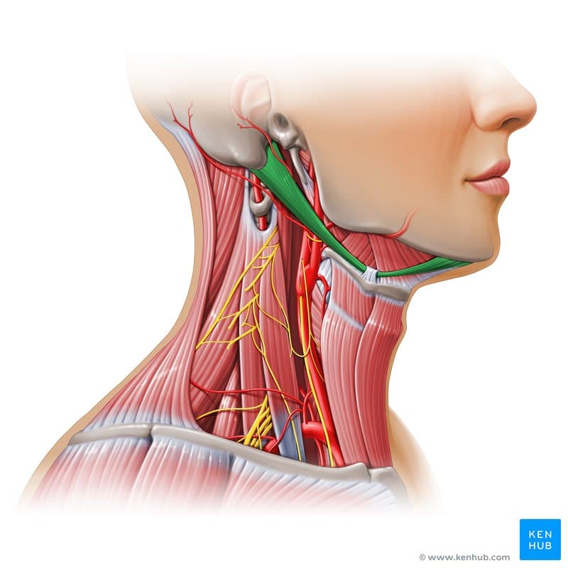
The digastric is a two-bellied muscle consisting of an anterior and a posterior belly, connected to the hyoid bone by an intermediate tendon.
Origin and insertion:
Innervation:
Functions:
This muscle plays a vital role in swallowing, speech, and movements of the lower jaw.
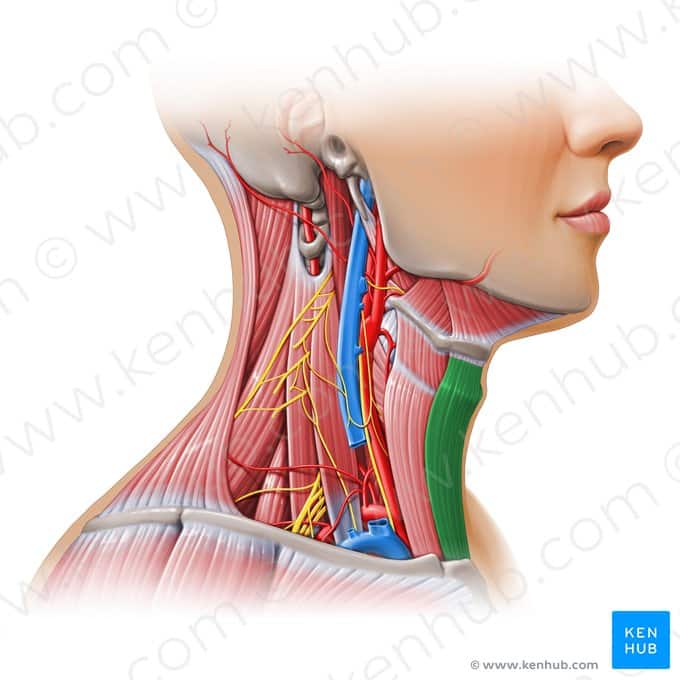
The sternohyoid is a long, superficial muscle in the neck that originates from the sternum and attaches to the hyoid bone.
Innervation: by branches of the cervical nerves (C1 to C3) through the ansa cervicalis.
Functions:
This muscle is part of the infrahyoid muscles and plays a role in positioning the hyoid bone and assisting swallowing movements.
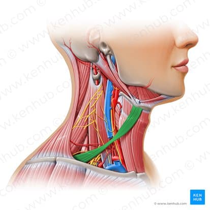
The omohyoid is a long, thin muscle from the infrahyoid group that consists of two parts (superior and inferior bellies) connected by an intermediate tendon.
Origin and insertion:
Innervation: by cervical nerve branches (C1 to C3) via the ansa cervicalis.
Functions:
This muscle contributes to the balance and fine movements of the neck and hyoid bone.
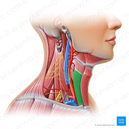
The sternothyroid is a long, deep muscle in the infrahyoid group that originates from the sternum and attaches to the thyroid cartilage.
Innervation: by cervical nerve branches (C1 to C3) via the ansa cervicalis.
Functions:
This muscle plays an important role in the movement and stability of the larynx and works in coordination with other infrahyoid muscles.
The thyrohyoid is a short, deep muscle in the infrahyoid group that originates from the thyroid cartilage and attaches to the hyoid bone.
Innervation: by the C1 nerve branch, which is carried via the hypoglossal nerve (cranial nerve XII).
Functions:
This muscle plays a key role in regulating laryngeal movements and stabilizing the hyoid bone.
Regular exercise strengthens the neck muscles, enabling them to better support the weight of the head and protect sensitive structures such as vertebrae, discs, and nerves. This added strength and endurance help prevent fatigue and injuries caused by repetitive movements or sudden stresses.
Exercise movements help stretch and increase the flexibility of neck muscles. This leads to improved range of motion of the head and neck and prevents movement limitations caused by muscle stiffness or tightness.
Strengthening and stretching exercises can reduce muscle tension and relieve excessive pressure on the cervical vertebrae and discs. This is especially beneficial for individuals experiencing neck pain caused by poor posture or repetitive activities.
Neck exercises help stabilize and strengthen the deep muscles that play a crucial role in maintaining balance and alignment of the cervical spine. This can lead to improved posture and prevent issues such as forward head posture or rounded shoulders.
The neck muscles play a vital role in head stability and overall body balance. Exercises targeting this area improve coordination between the neck, shoulder, and spinal muscles, reducing the risk of injury—especially for athletes and individuals engaged in intense physical activities.
Neck stretching exercises can relieve tension and pressure in the muscles of this area, promoting a sense of relaxation and comfort. This helps reduce both physical and mental stress.
Neck exercises and movements are not only essential for increasing the strength and function of neck muscles but also contribute to improving quality of life, reducing injuries, and preventing common neck problems. Performing these exercises regularly and with proper technique significantly enhances their positive effects.
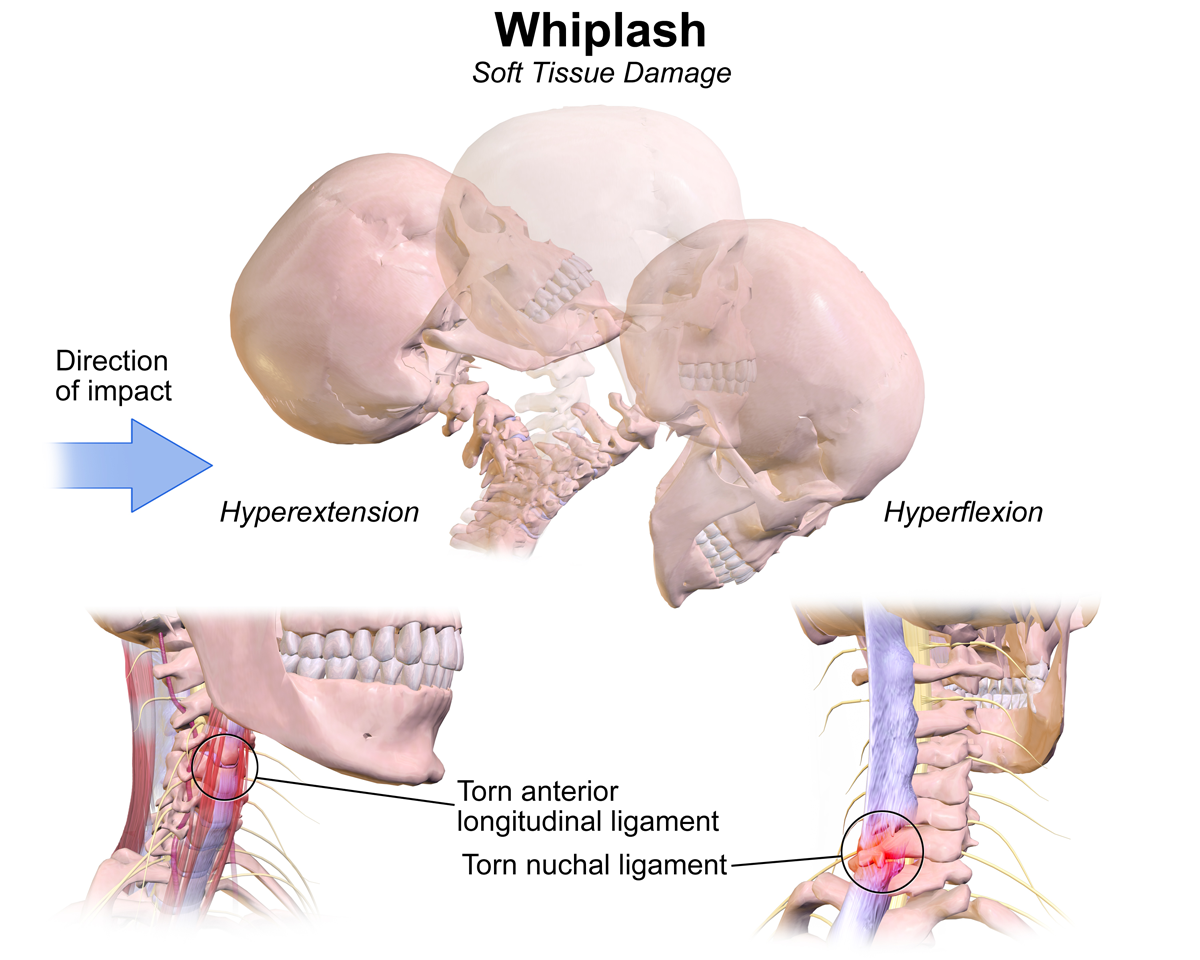
Whiplash is a neck injury that typically occurs due to a sudden, forceful movement of the head forward and then backward. This injury is commonly seen in car accidents, especially rear-end collisions.
Characteristics:
Treatment:
Whiplash typically improves with appropriate treatment, but in some cases, it can lead to long-term complications.
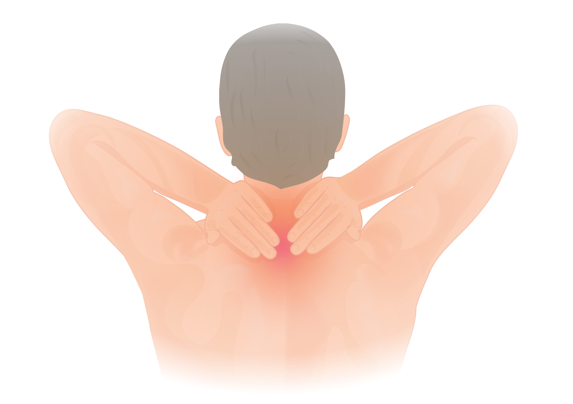
Neck muscle spasm is a condition in which the neck muscles contract suddenly or continuously, causing pain, limited movement, and sometimes headaches.
Common causes:
Treatment:
Prevention is possible by maintaining proper posture and performing neck exercises.
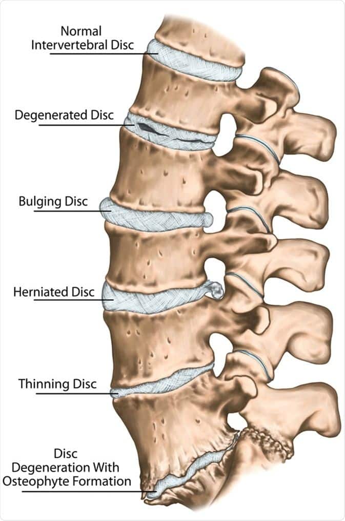
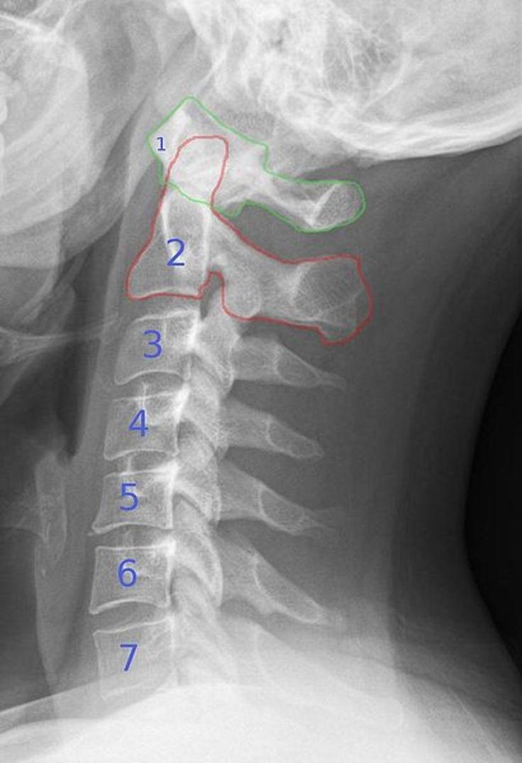
The cervical disc refers to the intervertebral discs in the neck, located between the cervical vertebrae (C1 to C7). These discs consist of an outer fibrous ring (annulus fibrosus) and an inner gel-like core (nucleus pulposus), which help absorb shock and maintain neck flexibility.
Common cervical disc problems:
Symptoms:
Treatment:
Ranges from rest and physical therapy to surgery in severe cases, depending on the severity of the condition.
Cervical Osteoarthritis or Cervical Spondylosis refers to the degeneration and wear of the joints, discs, and bones of the cervical spine. This condition usually occurs due to aging or gradual wear and tear of the vertebrae and discs.
Symptoms:
Causes:
Treatment:
This condition is usually chronic, but with proper care, the symptoms can be managed.
Physical therapy: Focuses on corrective and strengthening exercises to restore normal muscle function.
Massage therapy: This method helps reduce muscle tension, increase blood circulation, and improve flexibility.
Therapeutic injections: Such as corticosteroid injections to reduce inflammation in severe cases.
Use of braces: Cervical braces help stabilize the neck and reduce excessive movement during injury.
Rehabilitation exercises: These exercises are designed to gradually restore muscle function.
The neck muscles play a key role in maintaining body function, preventing injuries, and improving quality of life. Given the complexity and importance of these muscles, understanding their structure, function, and care methods can enhance both quality of life and athletic performance. Regular exercise, maintaining proper posture, and injury prevention are among the most important measures to strengthen these vital muscles.
Anatomy and medical books :
Gray's Anatomy (one of the standard references in anatomy)
Netter's Atlas of Human Anatomy (a well-known illustrated atlas in anatomy)
Clinically Oriented Anatomy by Keith Moore
Sports and training references :
Strength Training Anatomy by Frederic Delavier
Essentials of Strength Training and Conditioning by NSCA
Well-known articles and training programs by international coaches
Medical databases :
PubMed (for scientific and research articles)
MedlinePlus (health and medical information)
WebMD (for practical and general health information)
Specialized sports and health websites :
Images used:
(Kenhub) kenhub.com
The Best Body Health Calculators Using Scientific Methods
Developed by Pelank Life ©